46 |
Klinisk Biokemi i Norden · 4 2013
D (disability)
Coma
Focal or
diffuse
neurological
impairment
Hypoglycemia
Hyperglycemia
Hyperosmolarity
Intracranial pathology
(bleeding, ischemia)
Epilepsy/Seizures
Intoxication
Sinus-vein thrombosis
Severe anemia
Electrolyte disorder
Infection – sepsis
Infection – meningitis/
encephalitis
Liver failure
Eclampsy
Trauma
Cerebral oedema
Wernicke-Korsakoff’s
Malaria
Glucose
Osmolarity
ABG + lactate
Anion gap
Na, K, ionCa
Creatinine, Urea
Chloride
CRP, WBC diff
D-Dimer
INR, aPTT
ASAT, ALAT
Hb
TPK
MCV
HCG
Tox-screen
(urinary dipstick)
Ethylenglycol
Ethanol
Methanol
Paracetamol
B-SR
Malaquick
Myoglobin
Mg
Phosphate
Lupus anticoagulans
Valproate
Phenobarbital
E (exposure)
Temperature
Further signs
of disease
(rashes)
Hypothermia
Hyperthermia
Petechiae
TSH, T3, T4
Na, K, Creatinine
INR, aPTT
TPK, Hb
Coomb’s
Myoglobin
Homocysteine
Folat
Cobalamines
S-electrophoresis
Table 1.
The ABCD approach.
Cardiac arrest
Cardiac arrest is an immediately life-threatening
symptom, and must be addressed as soon as possible
as the duration of untreated cardiac arrest correlates
directly with mortality and long-term neurologic out-
come. The treatment of CA follows the Ilcor-guidelines
recommended by the ACC and the ERC, and focuses
on the presentation of CA as ventricular fibrillation
(VF) and non-ventricular fibrillation, divided into pul-
seless electric activity (PEA) and asystole. Even if the
cause of CA has been shown to be myocardial infarc-
tion in the majority of cases, the outcome of patients
with VF has been superior to the non-VF patients in
most of the studies.
This has been contributed to the fact that VF as the
cause of insufficient circulation could be diagnosed by
the ECG, and immediately addressed by the means of
electric conversion. However, there are treatable causes
of PEA/asystole, too, even if these require other investi-
gations to be performed during ongoing resuscitation.
These causes can be divided into 4 H’s (hypoxia, hypo-
volemia, hypothermia, hyper-/hypopotassemia and
electrolyte disorders) and 4 T’s tension pneumothorax,
cardiac tamponade, thrombembolism/PE, toxic). Table
2 shows which investigations to be performed during
resuscitation help to distinguish between these patho-
logies, and which further samples might be of interest
for further treatment.
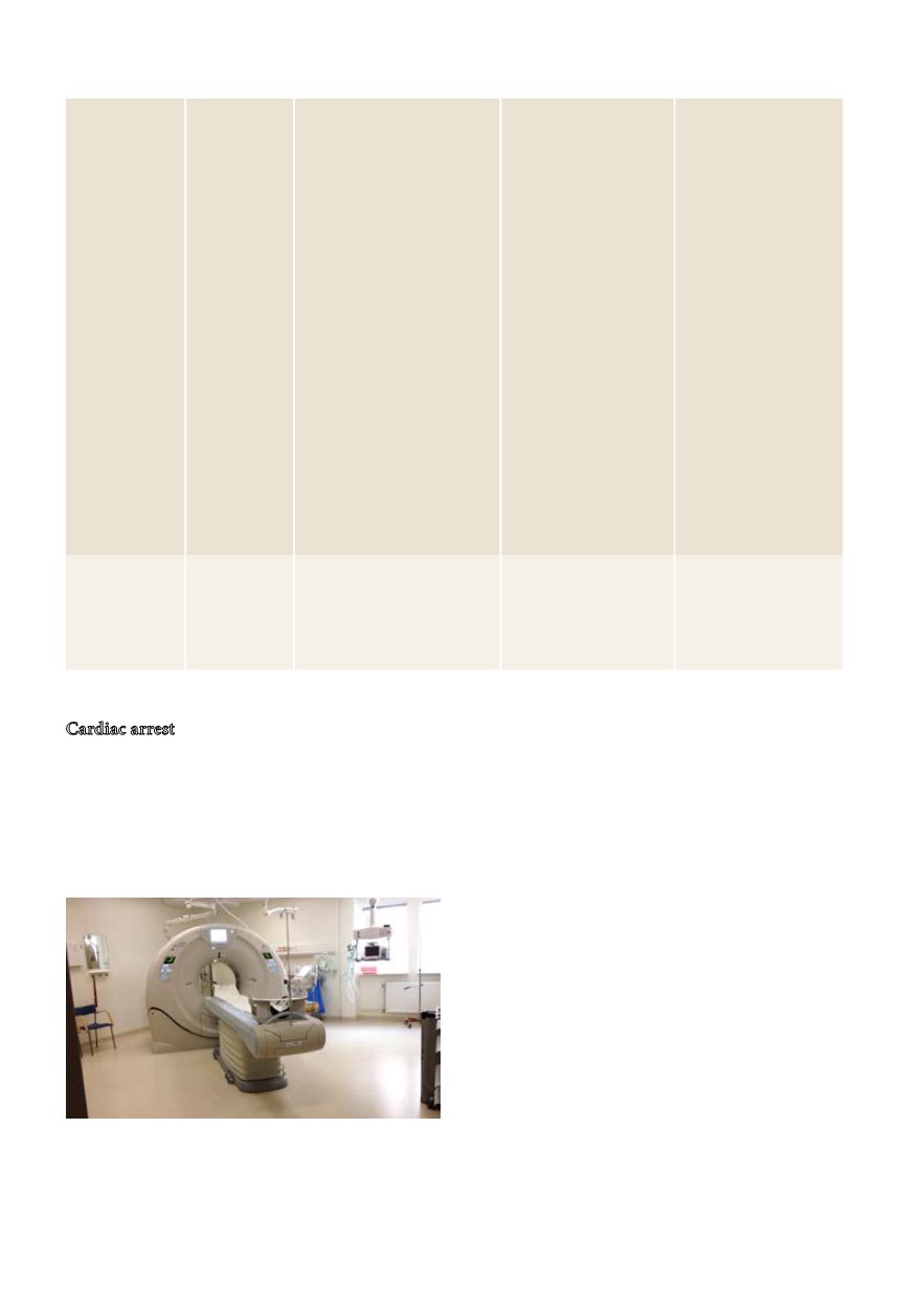
CT utförs t.ex. vid misstanke om stroke och kräver kreatinin
inom 5 minut (kontrastmedel) och PK (INR) inom 20 minut
(trombolys). (Foto: Per Simonsson).


